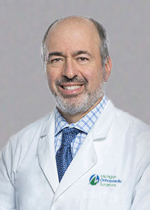
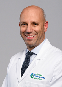
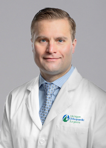
(March 13-16, 2019) The American Academy of Orthopaedic Surgeons hosted their Annual Meeting in Las Vegas, NV. Orthopaedic surgeons from around the world share their research and experience at the Annual Meeting to deliver our flagship education. The offerings include over 250 sessions in a variety of learning styles. Several MOS physicians participated and presented their work. See below to read more about each MOS doctor’s presentation and to learn more about each doctor in attendance.
Podium Presentations
Summary: With health care utilization becoming an important factor in patient care, we investigated the effect that surgical day of week has on patients’ length of hospital stay (LOS). We hypothesize patients having surgery on Friday will have a longer LOS than either Monday or Wednesday patients, especially for those being discharged to extended care facilities (ECF), due to decreased health care resources available on the weekend. All patients undergoing primary anatomic or reverse total shoulder arthroplasty by a single surgeon on Monday, Wednesday, or Friday between June 2006 and December 2016 were retrospectively reviewed. Revision surgery, outpatient surgery, and patients admitted through the emergency department were excluded. 1784 patients met the inclusion criteria Surgical day of week had a significant impact on LOS for patients undergoing shoulder arthroplasty. For those discharged home the mean LOS was 2.6 ± 1.3 days versus 4.3 ± 3.3 days for those discharged to ECFs. The percentage of patients discharged on each postoperative day (POD) shows that the majority of patients discharged home left on postoperative day (POD) 2 (58%), and those going to an ECF had the majority of discharges on POD 3 (58%) (Table 1). Day of surgery was found to have a more significant impact on LOS in patients being discharged to an ECF versus home. Patients discharged to ECF with Friday surgery had a significantly longer LOS than both Monday (P=0.028) and Wednesday (P=0.010) patients (Table 2). This effect was most evident in the subset of patients being discharged to an ECF. We believe the biggest contributor is the decreased healthcare personnel staffing over the weekend, since discharge to an ECF requires the coordination of many parties (physical therapists, care managers, insurance companies). These findings should be considered during surgical scheduling in order to minimize health care expenditures. Those patients who are at high risk for requiring ECF at discharge should be scheduled at the beginning of the week while more resources are available to expedite their discharge.
Summary: Modular total hip arthroplasty (THA) systems provide surgeons intraoperative options, but the taper-head interface is prone to fretting and corrosion. Previous studies have not clearly defined the cause of fretting and corrosion at this interface; however, ceramic-on-polyethylene (CoP) implants have exhibited lower fretting and corrosion scores than metal-on-polyethylene (MoP) implants. This study aims to investigate the effect of taper design on taper corrosion and fretting in modular CoP THA systems, as Wight et al. indicated in a recent meta-analysis of 91 studies focusing on reducing fretting and corrosion damage at the head-neck taper, that while the use of ceramic femoral heads was recommended at a “fair” level, evidence-based implant selection regarding taper geometry was “largely conflicting or inconclusive”. From 2002-2017, 120 retrieved CoP THA systems with zirconia toughened alumina (ZTA) femoral heads, with four different taper designs (11/13, 12/14, 16/18, V40), were identified. Femoral stem trunnions were visually evaluated and graded for taper fretting and corrosion using the Goldberg classification system as well as presence or absence of eight other damage modes of all components. There were no statistically significant demographic differences between taper groups for duration of implantation, laterality, patient age, and patient sex; however, patients with 16/18 tapers had a higher BMI than patients in the V40 taper cohort (p=0.012). In the study population, duration of implantation had a weak, positive correlation with both trunnion fretting (ρ=0.224, p=0.016) and corrosion (ρ=0.253, p=0.006); specifically, in the V40 group, there was a moderate, positive correlation between duration of implantation and summed corrosion score (ρ=0.443, p=0.021). In the V40 group, there was also a moderate, positive correlation between BMI and summed fretting score (ρ=0.458, p=0.014). Summed fretting and corrosion scores were significantly greater on the V40 and 16/18 tapers compared with the 12/14 tapers (all p≤0.001; Figure 1). Moderate-to-severe fretting damage was greatest in the V40 and 16/18 taper cohorts, while moderate -to- severe corrosion damage was greatest in the 16/18 and 11/13 groups (Figure 1). In the 11/13 group, there was a moderate, positive correlation between scratching of the femoral head and summed corrosion score (ρ=0.491, p=0.037). In the 120-explant population, 67 explants (56%) had the same grade across all four taper regions (superior, posterior, anterior, inferior quadrants), the full circumference of the taper, for both fretting and corrosion. Matching grades of the taper circumference were observed in 83 (70%) trunnions for fretting alone and 82 (69%) trunnions for corrosion alone. Of the 67 explants with overall identical grades for both fretting and corrosion, 55 (82%) trunnions had no damage (grade=1), 10 (15%) had mild damage (grade=2), and 2 (3%) had severe damage (grade=4). There were no explants that had matching grades of moderate damage (grade=3) across all four regions. Summed fretting and corrosion scores were greatest in the V40 and 16/18 taper groups and lowest in the 12/14 taper group. Differences in taper design characteristics may lead to greater micromotion at the taper-head interface, leading to increased fretting and corrosion. A generous gift provided by Byron and Dorothy Gerson to establish the Byron and Dorothy Gerson Implant Analysis Fund has supported this research.
Summary: Postoperative dysphagia following anterior discectomy and fusion (ACDF) is a multifactorial issue, but is strongly correlated with paravertebral soft tissue swelling at the surgical level. Previous research by our group demonstrated that implanting an absorbable gelatin (GelFoam, Baxter Biosurgery) sponge loaded with methylprednisolone in the retropharyngeal space at the time of ACDF is associated with a lower incidence of postoperative dysphagia. Concerns remain, however, that the potent anti-inflammatory effects observed with local steroid delivery may negatively impact new bone formation and ultimately fusion. Under an IRB-approved protocol, a retrospective analysis of 121 patients that undergone ACDF with a cortical allograft packed with demineralized bone matrix along with anterior plate and screw fixation was performed. The intervention group consisted of patients (n=42) who had an absorbable, steroid-loaded (80 mg methylprednisolone) implanted in the retropharyngeal space at the time of ACDF. The control group consisted of matched cases based on number of spinal levels treated and age at an approximately 1:2 case to control ratio (n = 79). 34 patients in the experimental group (80.95%) and 55 patients in the control group (69.62%) had sufficient radiographic follow-up to be included in the radiographic analysis. There was no significant difference between case and control groups in terms of sex (57.14% female vs 45.57% female, respectively, p=0.2254). Steroid and nonsteroid groups had similar BMI (29.69 vs 29.9, respectively, p=0.851). The steroid group trended towards a higher rate of diabetes mellitus (23.81% vs 11.39%, p=0.0735), but a lower ASA score (69.05% with ASA of 1 or 2, versus 51.90%, p=0.0688), although neither of these reached statistical significance. The steroid group had a longer operative time (97.55 vs 83.47 min, p 0.0056), as well as higher blood loss (31.79 mL vs 16.71 mL, p=0.0001). 34 patients in the experimental group (80.95%) and 55 patients in the control group (69.62%) had sufficient radiographic follow-up to assessed for radiographic fusion. The steroid group had a fusion rate of 64.7%, while the control group had a fusion rate of 91% (p=0.005). When analyzed by attempted fusion levels, the steroid group had a patient fusion rate of 81%, while the control group had a rate of 93% (p=0.025). One patient in the steroid group developed an esophageal rupture and retropharyngeal abscess, requiring surgical irrigation, debridement, and repair 8 months postoperatively. There was no difference in all cause re-operation (3/42 in steroid group, 9/79 in control group, p= 0.5397). There was no difference in reoperation for pseudarthrosis (1/42 in steroid group, 4/79 in control group, p= 0.6574). Given the differing reported efficacy of retropharyngeal steroids in prospective, randomized controlled trials, as well lower rates of radiographic arthrodesis in our series, we do not recommend the use of local steroids after ACDF. Future studies should examine larger groups in a prospective setting, and should include the use of clinical outcome measures.
Summary: Adjacent segment disease (ASD) is the most common cause for revision after anterior cervical discectomy and fusion (ACDF). This conditions is characterized by degenerative changes to the motion segment immediately adjacent to a level that has undergone a fusion surgery. Recently, stand-alone cages (SAC) have been developed for ACDF procedures. These interbody fusion cages have integrated screws for fixation, obviating the need for additional plate and screw fixation. In the setting of ASD, employing SACs reduces or eliminates the need to remove hardware from previous surgeries, which may reduce operative times and blood loss during surgery. While SACs have been compared to traditional hardware in the setting of primary fusion, there is a lack of data on radiographic fusion outcomes when SACs are utilized during ACDF performed to address ASD. Under an IRB-approved protocol, a retrospective chart and radiographic analysis was conducted to compare surgical outcomes and radiographic fusion rates in patients that underwent ACDF for a primary diagnosis of ASD using either a SAC (n=xx), or a traditional anterior plate construct (APC, n=xx) with screws. An additional cohort of patients who underwent primary ACDF for atraumatic and non-neoplastic diagnoses with traditional APC and screws was included to provide further context of fusion rates. Radiographic fusion was assessed on flexion-extension X-rays and defined as flowing bone in the interbody space with less than 2 mm of motion between posterior elements, as described by Cannada, et al. Patients underwent standard of care computed tomography (CT) scans at one year post-op. The rate of fusion for primary ACDF was higher than the fusion rate for all patients undergoing surgery for symptomatic ASD (95% vs 73.91%, p=0.009). ASD patients trended towards higher reoperation rates, although this did not reach statistical significance (9.52% vs 0%, p=0.1199). The fusion rate for primary ACDF was higher than the fusion rate for patients undergoing surgery for symptomatic ASD with APC, although this did not reach statistical significance (95% vs 82.35%, p=0.1509). ASD SAC patients had a significantly lower fusion rate compared to the primary ACDF patients (68.97% vs 95%, p=0.0060), as well as a significantly higher reoperation rate (13.79% vs 0%, p=0.0275). ASD APC and ASD SAC had no significant difference in fusion rates (82.35% vs 68.97%, p=0.4893) or reoperation rates (0% vs 13.79%, p=0.2810). There were 4 reoperations, all in the ASD SAC group. Two patients had hardware migration require re-operation, and two patients required surgery for pseudarthrosis. In conclusion, symptomatic ASD can be managed surgically with either revision anterior plating, or a stand-alone (i.e. zero-profile) cage. Although SACs have may have benefits in terms of dysphagia and ease of implantation in the revision setting, in our study, we found a lower rate of radiographic fusion as well as a higher rate of reoperation in the setting of ASD compared to a primary single-level ACDF with APC. We also saw a lower rate of fusion for ASD, regardless of implant type, compared to ACDF in the primary setting. Although much of the literature favors the use of stand-alone cages in anterior cervical arthrodesis, differences between device design and rigidity may have real implications in fusion rate. Future studies should focus on larger sample sizes with prospective designs, as well as a focus on clinical outcome measures.
Summary: Reverse total shoulder arthroplasty (RTSA) has become a common treatment for comminuted proximal humerus fractures (PHFs) and failure of open reduction internal fixation (ORIF) for PHFs in elderly patients. However, most published outcome data are for older-generation, cemented components. Our objective was to determine if current-generation, uncemented implants would provide similar outcomes while avoiding the complications associated with cement. We reviewed patients that underwent uncemented RTSA by two shoulder surgeons as initial treatment for PHFs as well as patients who underwent uncemented RTSA status post failed ORIF for PHF between 2008 and 2015 at our institution. 30 patients that had RTSA for PHF had adequate follow-up and were included in the study. 20 patients that had RTSA for failed ORIF had adequate follow-up and were include in the study. Patients that had RTSA immediately following PHF had better ASES (American shoulder and elbow surgeon) shoulder function score postoperatively than those that had RTSA following failed ORIF (82.0 ± 13.5 vs 64.0 ± 27.2, P=0.016). Postoperative pain scores did not vary between groups (P=0.301). Patients that had RTSA immediately following PHF had greater forward elevation than those that had RTSA following failed ORIF (130 ± 31 vs 107 ± 31, P=0.035). There was greater healing of tuberosities in patients that had ORIF (100% healing rate) than those that went straight to RTSA (70% healing rate). The rate of humeral stem subsidence, radiolucent zones, and heterotopic ossification was similar in each group. In conclusion, patients that have a RTSA immediately following PHF may have better outcomes than those that have ORIF and are converted to RTSA down the line.
Poster Presentations
Summary: In this study, the rate of VTE in the postoperative setting of revision total joint arthroplasty for periprosthetic joint infection (PJI; only cases that met MSIS criteria were included) was investigated. Efficacy of different anticoagulation regimens was also assessed. In the 5-year study period, 232 cases of revision TJA for PJI were identified. Low-dose aspirin was used in 14 cases, high-dose aspirin was used in 87 cases, and other anticoagulation treatments were used in 131 cases. Of the 232 patients, 9% (n = 21) had a prior history of DVT, none of whom developed a new VTE during chemoprophylaxis treatment. The incidence of VTE after revision surgery was 6% (n=15; DVT=11, PE=4). VTE rates were not significantly different between the three anticoagulation regimen groups (p=0.835). When the aspirin groups were combined, VTE rates between aspirin and other treatments was not significant (p=0.642). Among culture-positive cases, there was a weak, positive correlation between VTE (DVT and/or PE) and postoperative length of stay (ρ=0.267, p<0.001); similarly, in culture-negative cases, there was a moderate, positive association between prior history of a DVT and postoperative length of stay (ρ=0.439, p=0.018). In this study population, both low-dose and high-dose aspirin demonstrated similar rates of VTE, compared to other anticoagulants, indicating that aspirin may be an effective form of chemoprophylaxis after revision arthroplasty for PJI. [/av_toggle] [av_toggle title=’Antibacterial and osseointegrative efficacy of titania nanotube surfaces (MOS: Dr. Paul Fortin)‘ tags=” av_uid=’av-zkc3xg’]
Antibacterial and osseointegrative efficacy of titania nanotube surfaces (MOS: Dr. Paul Fortin)
Summary: When periprosthetic infection (PJI) often occurs in patients who have undergone orthopaedic procedures, the complication can substantially increase patient care costs; specifically, PJI may lead to more clinic/hospital visits, multiple revision surgeries, long-term patient disability, and increased mortality. Previous studies have shown that titania nanotube (TiNT) surfaces demonstrate increased bone-implant contact (BIC), enhanced de novo bone formation, and greater implant pull-out forces than non-textured controls. This study evaluated in vitro antibacterial properties of TiNT surfaces, TiNT surfaces integrated with nanosilver (TiNT+Ag), and two current standard-of-care materials (titanium thermal plasma sprayed and titanium alloy surfaces; TPS and Ti, respectively). Following in vitro testing, New Zealand White rabbits received bilateral antegrade implantation of intramedullary tibial implants with one of the four surface types. One tibia received a human clinical isolate of MRSA (105 CFU/mL) suspended in LB broth on the implant surface at the time of implantation (experimental limb), while the contralateral tibia received LB broth (no MRSA) (control limb). In a follow-on pilot study, a “therapeutic” cohort (TC) of rabbits underwent the same procedure, allowed infection to develop for 4 days, and then received vancomycin treatment (30 mg/kg; subcutaneous, twice per day) for 7 days. In vitro studies showed that when MRSA bacteria was directly seeded onto implant surfaces, there were lower MRSA counts on the TiNT groups at all timepoints and on the TiNT+Ag group at the 24 hr and 48 hr timepoints. TiNT surfaces with 110nm diameters showed the lowest bacteria counts compared to other nanotube diameters and were subsequently used for all in vivo work. In the in vivo experiment, sonication analysis showed that viable MRSA was greater on the TiNT and TiNT+Ag surfaces (104 CFU/mL, on average) vs. the Ti and TPS surfaces (103 CFU/mL, on average). In the TC, viable bacteria in the sonicant was approximately 102 to 103 CFU/mL for TiNT+Ag and TiNT implants, compared to 104 CFU/mL in the TPS group. μCT analysis of infected limbs showed greatest BIC on TPS implants, followed by TiNT, TiNT+Ag, and Ti. Bone-implant contact (BIC) was significantly greater in TiNT+Ag vs. Ti implants (p=0.023) and TPS vs. TiNT+Ag implants (p=0.031). In the TC, BIC% was greatest in the TPS group (0.277%) and approximately equivalent in the Ti, TiNT, and TiNT+Ag groups (0.15%, 0.1%, and 0.086%, respectively). Analysis of histologic sections demonstrated TiNT+Ag had the greatest BIC%, on average, (41%, range, 18-60), followed by TiNT (33%; range, 12-53), Ti (15%; range, 0-34), and TPS (12%, range, 3-28). In the TC, BIC% was greatest in TiNT+Ag implants (24%), followed by TPS (16%), Ti (15%), and TiNT (8%). On average, Ti had the lowest GP% (6%; range, 2-13), followed by TiNT+Ag (9%, range 3-14), TPS (10%, range 3-21), and TiNT (13%, range 1-26). In the TC, Ti had the lowest GP% (3%), followed by TiNT (5%), TPS (8%), and TiNT+Ag (15%). Sonication analysis indicated that TPS and Ti implants had less viable bacteria; however, overnight incubations showed that the implants continued to grow bacteria after sonication, perhaps indicating that differential adhesion of bacteria or decreased biofilm formation of the various surfaces may have interfered with assessment of in vivo antimicrobial efficacy. Specifically, the authors hypothesize that during sonication, bacteria detach more readily from nanotube surfaces, resulting in increased viable MRSA in the sonicant of nanotube implants compared with sonicant from TPS and Ti implants. With a reduction in viable bacteria from nanotube surfaces following antibiotic treatment, MRSA clearance rates may improve with the NT surface modification. Titiania nanotube surfaces may provide improvement over current implant technologies by simultaneously enhancing osseointegration, especially in non-tapered applications (e.g. tibial trays, fusion cages), as well as antibacterial properties. Work was funded through the University of Michigan MTRAC for Life Sciences Innovation Hub.
Summary: Our group recently demonstrated that postoperative mobilization of mesenchymal stem cells (MSCs) from the marrow compartment and into peripheral blood via subcutaneous granulocyte-colony stimulating factor administration enhanced tendon-bone healing in a rat model of surgical repair of a full-thickness supraspinatus tendon tear. In this study, we hypothesized that combining postoperative stem cell mobilization with local delivery of a chemokine that stimulates MSC chemotaxis would enhance the surgical repair of a mid-substance Achilles tendon transection. Achilles tendon of 24 Lewis rats were frayed then transected; then, the tendon anastomosis was coated with a resorbable chitosan hydrogel with or without CCL5 (RANTES). At four weeks, RANTES-loaded chitosan hydrogel had worse (higher) modified-Bonar subscores with respect to tenocyte morphology, collagen orientation, and ground substance staining, though none of these comparisons were statistically significant (p = 0.068, p = 0.672, and p = 0.407, respectively). Animals that received subcutaneous AMD3100 alone, or in conjunction with RANTES-loaded chitosan hydrogel displayed the best (lowest) total modified Bonar scores, though these comparisons were also not statistically significant (p = 0.498). No significant differences were found with respect to CD163 (p = 0.132) and CD68 (p = 0.104) staining. Despite a lack of statistical significance, animals that received AMD3100 displayed a higher number of both macrophage subtypes. No differences were found with respect to Collagen III immunostaining (p = 0.808). The mobilization of endogenous marrow-derived MSCs into peripheral blood and subsequent recruitment via local chemokine delivery may be an efficacious way to enhance tendon repair without the need for ex vivo processing. Work was funded through the American Orthopaedic Foot and Ankle Society (AOFAS).
Summary: Thirty percent of Americans have low back pain (LBP) at any given time, leading to approximately 50 million physician visits in the U.S. annually. For patients with chronic low back pain (CLBP), the etiology can be difficult to diagnose and then treat using either current non-surgical therapies, with inherent patient adherence challenges, or surgical interventions, with associated long recovery times. An anatomic study by Antonacci et al., proposed that a portion of pain previously ascribed to the intervertebral disc actually emanates from the vertebral body endplate nociceptors which communicate to the central nervous system (CNS) through the basivertebral nerve (BVN).1 We report on the two-year outcome data of the treatment arm patients in the SMART trial, which studied radio-frequency (RF) abalation of the BVN to treat CLBP. Under an IRB-approved protocol a total of 128 patients, from the per-protocol treatment arm of the SMART trial, a double-blind sham-controlled randomized trial, were followed for up to 24 months. Oswestry Disability Index (ODI) and pain visual analog scoring (VAS) instruments were used to collect patient-reported outcomes at baseline, 2 wks., 6 wks., 3 mo., 6 mo., 12 mo., and 24 mo. ODI and VAS data were available on 106 and 104 patients at 24 months respectively. The mean baseline ODI in the treatment arm was 42.4, decreasing to 22.6 at 12 months and 18.8 at 24 months. The mean baseline VAS in the treatment arm was 6.73, decreasing to 3.96 at 12 months and 3.13 at 24 months. Using a paired t-test comparing the 12 and 24-month VAS observations, the mean score at 24 months was significantly improved compared to 12 months (3.80 vs. 3.13, p=0.004). The SMART trial demonstrates that BVN ablation is statistically and clinically superior to the sham intervention, and that the degree of ODI improvement is approximately one disability category.2 Longitudinal follow-up of these patients demonstrates that ablation of the BVN for the relief of CLBP is a durable procedure with continuing improvement in patient reported ODI and VAS scores at up to two years.
Specialty Day Presentations
Innovative techniques in the treatment of cavovarus deformity of the foot and ankle.
Discussed ankle joint preservation techniques as an alternative to ankle fusion or replacement.


